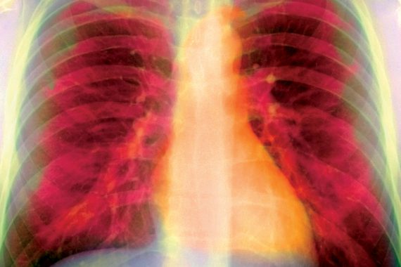

lungs and heart xray 3×2
Dear Dr Russell
Thank you for seeing this 42-year-old lady regarding a persistent cough and some shortness of breath in the context of abnormal focal signs in her lung.
The cough started about eight weeks ago, around the time of a mild URTI. She does not normally suffer from a cough and has no history of respiratory disease. The cough results in small amounts of clearish phlegm, with no blood. She feels moderately short of breath when she exerts herself, but she has no chest pains, and she otherwise appears to be keeping well with no weight loss or night sweats. She doesn’t smoke. She has not travelled abroad and she has no pets.
I have now examined her on three separate occasions, and each time I have noticed a patch of coarse crepitations at the base of her right lung. This has not shifted – and nor have her symptoms improved – with amoxicillin, erythromycin or doxycycline.
A chest X-ray was reported as normal and blood tests were fine other than a very marginally elevated CRP.
I’m at a loss to explain the persistence of her symptoms and signs. I’d be grateful for your thoughts on what the differential diagnosis might be here and how she should be investigated further.
Yours sincerely, GP
Dear GP
Thank you for asking me to see this lady. I believe we now have a way forward.
History
The patient has had an occasional cough in the past. She has not presented with these, but when pressed she reports they occur at least once a year, usually in winter. She also had a bout of pneumonia as a six-year-old and remembers being in hospital. This might point to an acute exacerbation of a chronic problem such as bronchiectasis. There are no clear red flags (weight loss, haemoptysis) but that does not fully exclude a potentially malignant process.
The patient says she is feeling tired but that this has come on slowly over the last year or so. She is still working part time, which is in many ways a good sign. Her symptoms seemed to start out of the blue and there does not seem to have been a significant infection, either viral or bacterial.
The patient denies any other relevant history, such as rheumatoid arthritis, lupus, chemotherapy or radiotherapy. Also, there was no evidence of gastro-oesophageal reflux disease or postnasal drip. She is currently not taking any cough-causing medication, such as an ACE inhibitor.
Examination
The patient has a widespread slight wheeze. This was soft and quite difficult to hear. There were coarse crackles heard best at the right lung base. These cleared with coughing to a degree. The finding that they are persistent and unilateral makes pulmonary fibrosis or other generalised lung disease less likely.
Investigations
The patient has a normal CXR. Spirometry showed mild airway obstruction. FEV1 was 1.65l (72% predicted), FVC 2.85 (102% predicted). The normal chest X-ray, in the context of no red flags, is reassuring in terms of cancer, especially in a non-smoker, but some studies have shown that up to 10% of chest X-rays in patients with lung cancer are reported as normal.
A slightly raised CRP is not of much help and implies minor systemic inflammation. If the CRP was significantly raised (>50) I would have expected a more focal history or signs of active infection. This is not the case. Differential diagnosis:
• Resolving pneumonia with post-infectious cough.
• Bronchiectasis (if evidence of previous episodes).
• Possible obstructive lesion.
• Possible underlying asthma.
Plan
• Full lung function tests were performed to investigate breathlessness, lung volumes and lung efficiency.
• CT scan of chest – a staging scan with high-resolution cuts
to look for bronchiectasis and exclude malignant process.
• I have given a low dose of inhaled steroids.
The CT scan revealed a focal area of bronchiectasis in the right middle lobe consistent with previous infection (perhaps in childhood). Lung function was normal with a good prognosis. She was referred for physiotherapy for chest clearance, and to be taught how to perform an active cycle of breathing and huffing to enable self-management.
Yours truly, Dr Richard Russell
Dr Richard Russell is a consultant chest physician at Lymington New Forest Hospital, Hampshire
Key points
A non-smoker presenting with persistent cough that eludes diagnosis should be asked about seasonal cough or childhood history
Other comon causes such as autoimmune diseases, GORD or drug therapy should be explored
A normal chest X-ray does not always exclude cancer and a high-resolution CT scan may be needed, which will also check for bronchiectasis
Full lung function testing and a trial of therapy can exclude underlying asthma
Pulse October survey
Take our July 2025 survey to potentially win £1.000 worth of tokens


