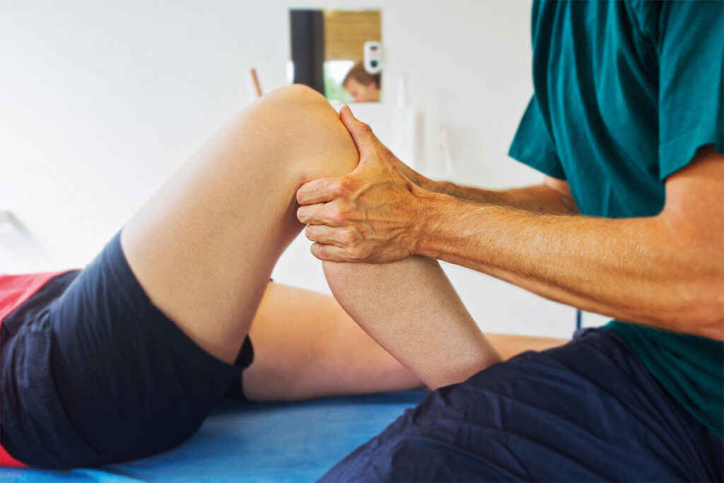
Managing sports injuries in the GP surgery: knee

Sports medicine specialists Dr Neil Heron, Dr Dáire Rooney and Dr Emma Gilmartin explain how to deal with knee injuries occuring through physical activity
Case 1: Medial meniscus injury
A 25-year-old female hockey player attends your morning clinic. ‘I twisted my knee last week on the pitch. It keeps catching and feels like it’s giving way when I walk on it.’
Typical presentation
Meniscal tears are a common injury both in athletes and in the general population, with the medial meniscus being the most commonly affected.1 Early diagnosis is essential to allow rehabilitation, thus increasing the likelihood of a positive outcome while reducing the risk of future osteoarthritis in the affected knee. They are similar to anterior cruciate ligament (ACL) injuries, in that the patient usually describes a twisting mechanism of injury.
Other key pointers are:
- Pain, typically along the medial or lateral joint line and exacerbated in weight-bearing activities such as squatting, or activities that involve rotation of the affected limb.
- The patient classically describes ‘locking’ or ‘catching’ in the affected knee. This sensation is usually due to an unstable tear or detached loose body in the knee. In extreme circumstances, the patient cannot extend the knee at all.
- Instability: the knee may be described as ‘giving way’.
- Swelling; this is typically slower in onset than with an isolated ACL injury and may not become apparent until three days after the injury. There may be recurrent knee swelling.
How to examine
Differentiating medial meniscus injury from an ACL injury is not always simple. Meniscal injuries often occur alongside ACL and medial collateral ligament injuries (together known as the ‘unhappy triad’) or patellar dislocations. A systematic approach is vital.
There may be significant swelling at the knee. When moving the knee joint, crepitus is often felt and the range of motion is often significantly decreased compared with the uninjured limb. For most patients, palpation of the medial joint line will be uncomfortable and this is the most sensitive clinical test for meniscal tears.
Other clinical tests that are not as sensitive but can support your diagnosis are:
McMurray Test: with the patient supine, hold the affected knee in full flexion. Provide a valgus stress and rotate the tibia externally. Then bring the knee into extension. Then, provide a varus stress to the knee and rotate the tibia internally. Rotating the tibia aims to catch the injured meniscus. The test is considered positive if the patient experiences pain, locking or clicking in the knee.
Apley grind test: with the patient prone, stabilise the femur using one hand. With the knee in 90˚ flexion, provide an axial load with your other hand through the patient’s heel. Then move the heel into external rotation (to test the medial meniscus) and internal rotation (to test the lateral meniscus). This test is considered positive if the grinding motion achieved by internal and external rotation causes discomfort in the knee.
Management
Management of medial meniscus tears depends on their stability. A referral to orthopaedics will be required for the knee that is giving way or locking, while other meniscal tears are usually managed conservatively with physiotherapy, helping to strengthen the surrounding muscles and keep the person as mobile as possible.
Case 2: Anterior knee pain caused by patellofemoral pain syndrome (PFPS)
A 45-year-old recreational runner comes to you with knee pain. She states that it has been present for the past few months: ‘I can’t remember injuring it, but now I can’t sit in church for more than half an hour.’
Typical presentation
Some 20-40% of MSK-related consultations in primary care relate to anterior knee pain.2
Common causes of anterior knee pain include:
- PFPS.
- Patellar tendinopathy.
- Prepatellar/infrapatellar bursitis.
- Fat pad impingement.
- Quadriceps tendinopathy.
PFPS refers to all peripatellar or retropatellar knee pain not caused by other pathology. Patients with this condition will usually have a structurally normal knee with the pain resulting as a consequence of a functional issue in the knee. This may be due to muscle weakness or imbalances but also to changes in exercise intensity.
Key pointers
Pain location – pain behind or surrounding the patella is likely to be related to the patellofemoral joint, whereas pain inferior to the patella might suggest a patellar tendinopathy or fat pad impingement. Patients with PFPS often describe this as ‘deep inside the knee’ and it tends to be ‘achey’ and diffuse. It is also useful to determine whether this pain is bilateral, in which case it is more likely to be due to PFPS.
Onset and exacerbating factors
Pain onset is often gradual but may flare after an acute traumatic episode. Pain is exacerbated by walking or running downhill. Classically, patients with PFPS also experience pain when sitting for a prolonged period with knee flexion. This is referred to ‘movie-goers’ sign’ in textbooks. Alternatively, if activities like jumping worsen knee pain, this may be suggestive of a patellar tendinopathy.
Associated symptoms
Patients with PFPS may describe a sensation that their knee is catching, clicking or feels like it may give way. This is unlike true instability, where the knee will give way, usually because of ligamentous or meniscal injuries. Instead the feeling of instability is caused by muscle weakness or inhibition secondary to pain. In this case, a thorough examination is needed to rule out more severe pathology.
How to examine
Given the array of primary causes, it is vital to use a systematic method of assessing the pain to guide further investigation and management.
Inspection of the knee is usually normal, but may reveal surrounding muscle wastage. Small effusions are common. Tenderness is experienced on palpation of the medial, lateral and inferior borders of the patella. Crepitus when moving the joint is common in the general population and is often not linked to a specific pathology. Knee range of motion is usually normal.
When assessing PFPS, functional tests are key. Ask the patient to do single-leg squats, looking for good pelvic alignment and whether the knee tracks over their foot as they squat. If the pelvis appears very unstable or the knee draws inwards, this may suggest a functional issue contributing to PFPS.
Clinical examination tools should also be used to rule out any structural issues in the knee that may contribute to the pain, especially if the patient describes any sensation of instability.
Management
This is largely through physiotherapy and management of the chronic pain.
Case 3: Lateral knee pain due to iliotibial band syndrome (ITBS)
A 34-year-old female presents with lateral knee pain when running. She explains the pain typically starts after running 4km and is worse when running downhill. She used to run a lot several years ago and never had any issues and now is frustrated by the limitation caused by this pain.
ITBS is a common condition characterised by lateral knee pain and tenderness on palpation over the lateral aspect of the knee. It is an overuse injury caused by friction between the iliotibial band and the lateral epicondyle of the femur producing inflammation and pain. It commonly occurs in runners and is associated with weakness in the hip abductor muscles or excessive foot pronation.3
Key pointers
Although ITBS is often thought of as a runner’s injury, it is also frequently seen in other sports including football, hockey and skiing. New runners are at increased risk as well as those who have suddenly changed the distance or frequency of their runs. There is also evidence that running with worn-out trainers or running excessive mileage can predispose to this injury. Symptoms are typically worse when running downhill.
Examination
The Ober test is most commonly used to diagnose ITBS pain. The patient should lie on their side with affected side up. Support and guide the affected leg backwards, towards the patient’s rear, and gently drop it down towards the table. A positive test should elicit the lateral knee pain the patient is experiencing.
Management
The vast majority of ITBS cases will resolve with conservative management alone.4 This includes a combination of rest, adequate stretching and modification of running habits.
Physiotherapy plays an important role in rehabilitation. Anti-inflammatory medication can be used short term. Surgical management of ITBS is rarely done and would be a last resort if symptoms are refractory.
Case 4: Anterior cruciate ligament (ACL) injury
A 24-year-old semi-professional footballer comes to you on a Monday morning following an injury sustained two days previously. ‘I was running with the ball and when I tried to change direction, I heard a loud pop. I tried to play on but couldn’t – what do you think it is, doc?’
Typical presentation
ACL injuries can have a devastating impact on an athlete’s sporting career and significant implications for anyone’s future capacity to exercise. The mechanism of injury involves a twisting motion of the affected leg, usually when jumping, changing direction or slowing down. Contrary to perceptions, 75% of ACL injuries involve minimal to no contact.5
Key pointers include:
- ‘Popping’ sound from knee
- Rapid swelling around knee joint within 1 hour after the injury
- Feeling that leg is ‘giving way’ and unstable following injury
- Sensation that something is ‘going out of place’ in the knee
- Inability to weight bear
Examination
Presentations can vary and many patients have very few symptoms, so clinical examination is vital to supplement the history. However, this can be difficult within the acute setting due to pain. Assessment of the ACL can be done via a number of tests and these should be used together to support your diagnosis:
- Lachman’s: the single most important test. Involves supporting the thigh with one hand, with the knee flexed to 20 degrees and attempting to move the tibia anteriorly with respect to the femur. An ACL injury exhibits less than usual defined endpoint and laxity of the ACL.
- Pivot shift: useful for detecting instability and knee function. Requires patient to be relaxed and able to fully extend the knee. Take the patient’s foot with one hand and flex the hip to 30 degrees whilst internally rotating the tibia, then apply a valgus stress to the leg whilst flexing the knee from a position of full extension. This should recreate the uncomfortable sensation felt when the initial injury occurred. FIFA has produced a video that shows how to perform this test.6
- Anterior drawer: Often said to be the least sensitive.7 Flex knee to 90 degrees and sit on the patient’s foot. Once again, pull the tibia anteriorly with respect to the femur. A lack of solid endpoint would raise your suspicion that the ACL is damaged.
Examination should also involve a close inspection of the leg – for example, to check for any gross effusion/muscle wastage) and examination of the surrounding structures (including the contralateral knee) to help rule out alternative diagnoses, such as a meniscal injury, as ACL injuries seldom occur in isolation.
Management
This would involve urgent referral to orthopaedics for imaging (typically MRI) to confirm the diagnosis and agree a management plan.
Case 5 – Baker’s cyst associated with knee osteoarthritis
A 63-year-old man presents with worsening pain in his right knee which he attributes to his ‘wear and tear’ arthritis. He is concerned as over the past two months he has felt a small lump at the back of his knee. He feels his symptoms have increased since this developed and that the lump has increased in size since running more on hard, road surfaces during his twice weekly 3-4km jogs.
Typical presentation
Baker’s cyst is an abnormal fluid distension of the gastrocnemio-semimembranosus bursa of the knee.8 In adults, Baker’s cysts are usually secondary to an underlying condition, most commonly osteoarthritis, inflammatory arthropathies, meniscal tears or damage to the anterior cruciate ligament. They are found in between 22-47% of patients with osteoarthritis of the knee.9 Adults can typically experience associated non-specific posterior knee pain, but this is often difficult to distinguish from the underlying cause.
Examination
Typical examination findings include a round, smooth and fluctuant bulge in the medial popliteal fossa. When in full knee extension, the cyst can feel firm and sometimes more difficult to palpate or softer when the knee is flexed – this is known as Foucher’s sign. This can be helpful to distinguish a Baker’s cyst from other popliteal masses which do not change with position. It is also important to be aware of potential complications including chronic pain, dissection or rupture, haemorrhage or DVT.
Management
Patients often ask for drainage of the Baker’s cyst but we tend to avoid this as they recur and there is a danger of damaging surrounding structures, including popliteal artery and vein. Obesity is often a factor and is recognised as a primary modifiable risk factor for osteoarthritis of the knee.10 Therefore, patient education on weight loss and the benefit of physical activity and exercise are important initial steps in effective management. A Cochrane review showed that the benefits of exercise outweigh those of simple analgesics.11 GPs should take time to educate their patients as it is a common misconception that exercise will exacerbate symptoms. It is also important to recognise that pain is a modifiable symptom influenced by many biopsychosocial factors of which many can be managed non-surgically.
There is of course a role for pharmacological management including adequate analgesia. First line treatment typically includes paracetamol and topical NSAIDs, with oral NSAIDs usually reserved for second line management. Referral to orthopaedics should be considered prior to the point of functional limitation, if symptoms are significantly affecting quality of life or if there is any uncertainty regarding the diagnosis.
Dr Neil Heron is a GP and consultant in sports medicine at Queen’s University, Belfast, and Dr Dáire Rooney and Dr Emma Gilmartin are FY2 doctors with an interest in sports medicine at the Royal Group of Hospitals, Belfast
References
- Clayton R, Court-Brown C. The epidemiology of musculoskeletal tendinous and ligamentous injuries. Injury 2008;39:1338-44
- van Middelkoop M, van Linschoten R, Berger M et al. Knee complaints seen in general practice: active sport participants versus non-sport participants. BMC Musculoskelet Disord 2008;9:36
- Taunton J, Ryan M, Clement D et al. A retrospective case-control analysis of 2002 running injuries. Br J Sports Med 2002;36:95-101
- Beals C, Flanigan D. A review of treatments for iliotibial band syndrome in the athletic population. J Sports Med 2013;2013:367169
- Boden B, Dean G, Feagin J Jr et al. Mechanisms of anterior cruciate ligament injury. Orthopedics 2000;23(6):573-8
- FIFA Medical Network. Pivot shift test. Anterior cruciate ligament rupture. Available at: https://www.youtube.com/watch?v=2TPfLOcxbTI [Accessed 1 June 2022]
- Katz J, Fingeroth R. The diagnostic accuracy of ruptures of the anterior cruciate ligament comparing the Lachman test, the anterior drawer sign, and the pivot shift test in acute and chronic knee injuries. Am J Sports Med 1986;14(1):88-91
- Mukund K, Subramaniam S. Skeletal muscle: A review of molecular structure and function, in health and disease. Wiley Interdiscip Rev Syst Biol Med 2020;12(1):e1462-e
- Naredo E, Cabero F, Palop MJ et al. Ultrasonographic findings in knee osteoarthritis: a comparative study with clinical and radiographic assessment. Osteoarthritis Cartilage 2005;13(7):568-574
- Lee R, Kean W. Obesity and knee osteoarthritis. Inflammopharmacology 2012;20(2):53-8
- Fransen M, McConnell S, Harmer A et al. Exercise for osteoarthritis of the knee: a Cochrane systematic review. Br J Sports Med 2015;49(24):1554-1557
Pulse July survey
Take our July 2025 survey to potentially win £1.000 worth of tokens

Visit Pulse Reference for details on 140 symptoms, including easily searchable symptoms and categories, offering you a free platform to check symptoms and receive potential diagnoses during consultations.
Related Articles
READERS' COMMENTS [1]
Please note, only GPs are permitted to add comments to articles


very good practical review