What to do with incidental kidney lesions
The finding of an incidental kidney lesion may result in anxiety and uncertainty for the patient and their clinician, raising concerns about the significance and immediate and future management.
Post-mortem studies suggest that at least 50% of people have at least one renal abnormality,1 so it is therefore not surprising that abnormalities on renal imaging are commonly reported. In primary care, abdominal ultrasound is the most common way the kidneys are imaged and this is the focus of this article.
Presentation
The kidneys may be imaged during any abdominal ultrasound scan. Patients may have presented with symptoms that are often unrelated to the renal tract. Right upper quadrant pain, abnormal liver function tests or non-specific ‘loin’ pain are common reasons for requesting abdominal ultrasound. Incidental abnormalities may also be detected on scans targeted at the renal tract, for example during the investigation of a new diagnosis of chronic kidney disease. Alternatively, a scan may have been done for completely unrelated reasons, such as obstetric ultrasound.
What abnormalities might be found?
The commonest abnormalities of the renal tract detected on ultrasound include:
- Renal cysts.
- Solid renal mass.
- Discrepancy in renal size.
- Solitary kidney.
- Abnormality of renal outline or irregularity of cortical thickness.
- Renal stone or other suspected calcification.
- Hydronephrosis.
- Duplex kidney.
Ignore
Renal cysts are common and are bilateral in 9% of people over the age of 70.2 Ultrasound is a reliable modality for visualising most renal cysts. Simple cysts have a characteristic appearance and are benign. The reporting clinician will add additional commentary if there is doubt about the nature of the cyst. In general, a single thin septum or small area of calcification within the cyst wall will not alter the presumption of benignity and no follow-up is required. Additional imaging will be required for more complex cysts, but this should be clear from an ultrasound report. Multiple cysts are more common with increasing age but can cause diagnostic confusion with a range of multi-cystic kidney diseases. The commonest of these is autosomal dominant polycystic kidney disease. The patient’s age and family history need to be considered to avoid missing this diagnosis (table 1).
Angiomyolipomas (AML) are benign tumours that may be identified by ultrasound. Most contain fatty elements. Although fatty AML can have a typical appearance, ultrasound is less reliable than other imaging modalities and confirmation with CT or MRI may be recommended. One practical approach is to perform additional imaging if the lesion does not have a typical ultrasound appearance or is 1cm or greater in size. A recommendation will often be made in the radiologist’s report. If the diagnosis of AML has been confirmed, solitary small lesions (<1 cm) are not clinically significant,3 providing there is no family history or clinical features of tuberous sclerosis.4
Larger AML may require follow-up, as lesions greater than 4cm have an increased risk of spontaneous haemorrhage and require intervention.
Duplex ureters are present in 1% of the population,5 although many will not be visualised by ultrasound. The majority are asymptomatic and of no clinical significance. In the absence of dilatation of one part of the collecting system or symptoms, such as recurrent urinary tract infection, they do not usually require further investigation.
Irregularity of the renal outline may be related to foetal lobulation, which is a normal variant. Scarring with irregular thinning of the renal cortex, however, suggests significant pathology. In younger patients this is most often associated with reflux nephropathy and consequent chronic pyelonephritis. In older patients, focal renal infarction should be considered, especially in the presence of atrial fibrillation.
| Age 15-29 | 30-59 | ≥60 | |
|---|---|---|---|
|
Known APKD1 gene |
≥2 cysts in total |
≥2 cysts in each kidney |
≥4 cysts in each kidney |
|
Age 15-39 |
40-59 |
≥60 |
|
|
Unknown genotype |
≥3 cysts in total |
≥2 cysts in each kidney |
≥4 cysts in each kidney |
Table 1
Monitor
Normal kidneys have a length of 10-14cm. A difference in length of ≥2cm on ultrasound is considered abnormal.6 A smaller discrepancy can be regarded as within the limits of normal variation. Reasons for a significant difference in renal size might include congenital renal dysplasia, chronic pyelonephritis secondary to reflux and ascending infection in childhood, renal infarction or unilateral renal artery stenosis or occlusion. Compensatory hypertrophy of the contralateral kidney implies that the abnormality within the smaller kidney arose in early life, most commonly due to unilateral renal dysplasia. If the person is asymptomatic, normotensive, has normal kidney function (eGFR), a negative urine dipstick for blood and a normal albumin to creatinine ratio (ACR), then monitoring in primary care with annual blood pressure, eGFR and urine ACR is appropriate. If the diagnosis is uncertain or if there is evidence of renal disease, referral to secondary care may be more appropriate.
Solitary kidney (unilateral renal agenesis) has an incidence of approximately one in 1,000.7 The implications for monitoring are the same as for dysplasia.
The incidental finding of renal stones in the periphery of the collecting system should lead to an exploration of risk factors for stone formation, including family history, dietary factors, fluid intake and a metabolic stone screen. Modifiable risk factors should be addressed.
Renal stones within the renal pelvis (including stag horn calculi) or ureter should lead to a referral to a urologist for more active intervention.
Refer
Complex cysts may be malignant and require referral for further investigation. This should be clear from an ultrasound report.
The majority of non-fatty solid kidney lesions are renal cell carcinoma. An incidental finding on a scan performed for other reasons is now the most common way for kidney cancer to present.
All such lesions should be referred for further assessment using a cancer referral pathway, unless there are good reasons to think that a conservative approach would be more appropriate. This might be the case in the presence of extensive comorbidity or a short life expectancy.
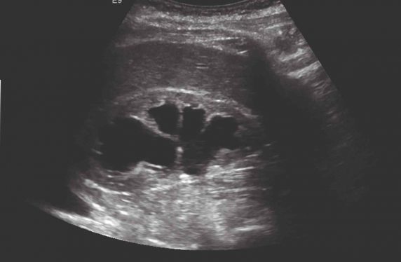
The finding of a unilateral or bilateral hydronephrosis raises the possibility of obstruction of the renal tract somewhere between the pelvi-ureteric junction and urethra. However, hydronephrosis is frequently overdiagnosed at ultrasound for two principle reasons. First, a failure to recognise the minor degree of pelvicalyceal distention that is normal in a well-hydrated patient and the frequent misrecognition of a normal anatomical variant, the extrarenal pelvis. Second, benign parapelvic cysts are misidentified as dilated calyces. An experienced sonographer or second opinion can usually distinguish between these situations, but additional investigation will sometimes be required to confirm there is no significant pathology.
True hydronephrosis leads to two important questions. First, is renal function normal? Bilateral obstruction or unilateral obstruction of a single functioning kidney will lead to severe kidney failure and requires urgent assessment. Second, what is the cause of the obstruction? While bladder outflow obstruction secondary to benign prostatic hyperplasia is the commonest cause of obstructive uropathy, malignancies intrinsic to the urinary tract (carcinoma of the prostate, bladder, ureter or renal pelvis) or extrinsic to the urinary tract (carcinoma of the cervix , rectum, retroperitoneal lymphoma and retroperitoneal metastases) are also considerations that need prompt investigation and exclusion.
Primary retroperitoneal fibrosis is a rare, but treatable cause of renal failure due to bilateral ureteric obstruction. Most patients with hydronephrosis therefore need urgent referral to secondary care.
Dr Colin Jones is a consultant renal physician and Dr Niall Warnock is a consultant radiologist at the York Teaching Hospital NHS Foundation Trust
Pulse July survey
Take our July 2025 survey to potentially win £1.000 worth of tokens

Visit Pulse Reference for details on 140 symptoms, including easily searchable symptoms and categories, offering you a free platform to check symptoms and receive potential diagnoses during consultations.


