How to recognise and diagnose primary cutaneous lymphomas

Under the radar: As part of our series discussing diagnoses that can potentially be overlooked in primary care, Dr Alice Eyers describes a case of primary cutaneous B-cell lymphoma that was initially treated as a fungal infection
Note details of this case have been changed to protect individuals’ identities
Clinical history
An 84-year-old man was discharged to a nursing home from hospital after a 4-week stay. The initial presentation was that of community acquired pneumonia, which was treated with intravenous antibiotics. His stay was prolonged by a slow recovery, reduced mobility and the presence of a rash.
The discharge summary noted that the rash was on the lower limbs and had been treated initially as a fungal infection with topical terbinafine. The rash failed to improve and was noted to have become slightly more raised. He had been discharged with a diagnosis of phlebitis, completing a further course of antibiotics with a request for the GP to review at the end of the course.
I visited him at home. He reported that he was slowly recovering but the rash had not improved and that it actually significantly predated his hospital admission. On examination, there were a number of violaceous nodules with no surrounding erythema or heat.
Making the diagnosis
The patient’s observations were within normal limits. Consent was sought from the patient and photographs were taken. These were then shared with the local dermatology team. The team organised an urgent appointment where biopsies were taken and a diagnosis of B-cell lymphoma was made.
The patient went on to have blood tests and a CT scan. These were unremarkable and the diagnosis was confirmed as primary cutaneous B-cell lymphoma. The patient underwent radiotherapy with a good response to treatment. He remains under surveillance follow-up.
What are primary cutaneous lymphomas?
These are non-Hodgkin lymphomas, confined to the skin. They are rare, easily missed and can often have a slowly progressive course, mimicking other conditions.
Peak incidence is between 50-74 years but 20% of cases are found in those aged 25-49 and children are also rarely affected. Advancing age is associated with poorer prognosis.
There are broadly two categories of cutaneous lymphomas. Primary cutaneous T-cell lymphomas account for 70% of cases, the most common of which is mycosis fungoides. Primary cutaneous B-cell lymphomas account for the remaining 30%.
An example of primary cutaneous B-cell lymphoma lesion as seen in this case is shown in image 1 below.
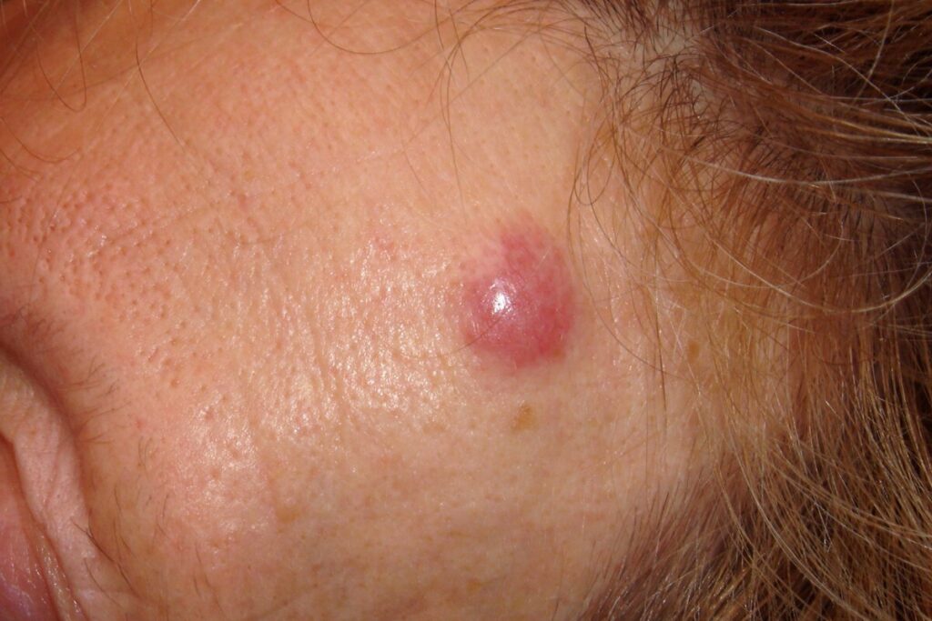
Image 1. Primary cutaneous B-cell lymphoma on the forehead of a 53-year-old woman
An example of a B-cell lymphoma that has progressed is shown in image 2.
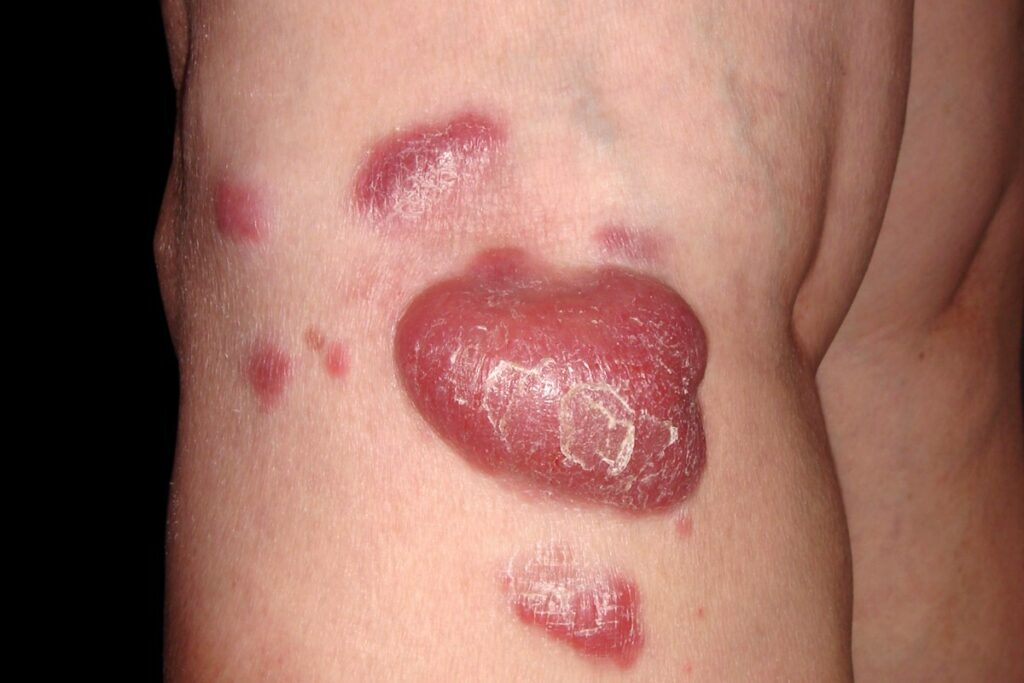
Image 2. Primary B-cell cutaneous lymphoma (diffuse large B cell lymphoma; leg type) on leg of 50-year-old woman
All patients found to have a cutaneous lymphoma need further investigation to ensure that there is not wider spread and that it is not in fact a cutaneous manifestation of a pre-existing systemic lymphoma. This is via imaging, specifically CT and PET scanning, and via specialist haematological examination of both blood and biopsy samples.
Treatment and prognosis
Topical treatments and phototherapy may be appropriate for early-stage mycosis fungoides. Other treatments options for higher stage mycosis fungoides and other types of cutaneous lymphomas include the use of biologics, radiotherapy and chemotherapy. Surgery can also be considered for solitary lesions.
Prognosis is dependent on the type of identified lymphoma and the stage. However, the prognosis for the most common type at stage I is good with a slow course and good response to treatment.
Learning points for GPs
- There are a variety of presentations from scaley patches to large nodules. Consider the diagnosis particularly in recurrent presentations or when there is an incomplete resolution of symptoms with potent topical steroids.
- Biopsy is difficult. Site of the biopsy is important and sometimes multiple biopsies are needed to get the diagnosis. If cutaneous lymphoma is suspected, a punch biopsy in surgery will not yield reassurance and referral to dermatology is needed.
- There is a link between some cutaneous lymphomas and Borrelia burgdorferi. All patients with a new diagnosis of cutaneous lymphoma should have serology checked and treatment with appropriate antibiotics if positive. With this in mind, a lower threshold for considering the diagnosis is prudent in patients with a known history of Lyme disease presenting with a new rash.
- As for other lymphomas, all patients with a new diagnosis of cutaneous lymphoma need to have an HIV test.
- British Association of Dermatologist guidelines on primary cutaneous lymphoma are currently being revised.
Dr Alice Eyers is a GP in Somerset
Sources/further reading
Gilson D et al. British Association of Dermatologists and UK Cutaneous Lymphoma Group guidelines for the management of primary cutaneous lymphomas 2018.Br J Dermatol 2019; 180(3: 496–526
Vermeer M. Epidemiology of cutaneous lymphoma. Br J Dermatol 2021;184(6):993–4
DermNet. Cutaneous B-cell lymphoma
Please note this article was updated on 6.03.26 to include example images
Visit Pulse Reference for details on 140 symptoms, including easily searchable symptoms and categories, offering you a free platform to check symptoms and receive potential diagnoses during consultations.
Related Articles
READERS' COMMENTS [1]
Please note, only GPs are permitted to add comments to articles


Pictures ??