Dealing with sports injuries in the GP surgery: foot and lower leg
Sports medicine specialists Dr Neil Heron, Dr Dáire Rooney and Dr Emma Gilmartin advise on how to deal with foot and lower leg injuries that occur through physical activity
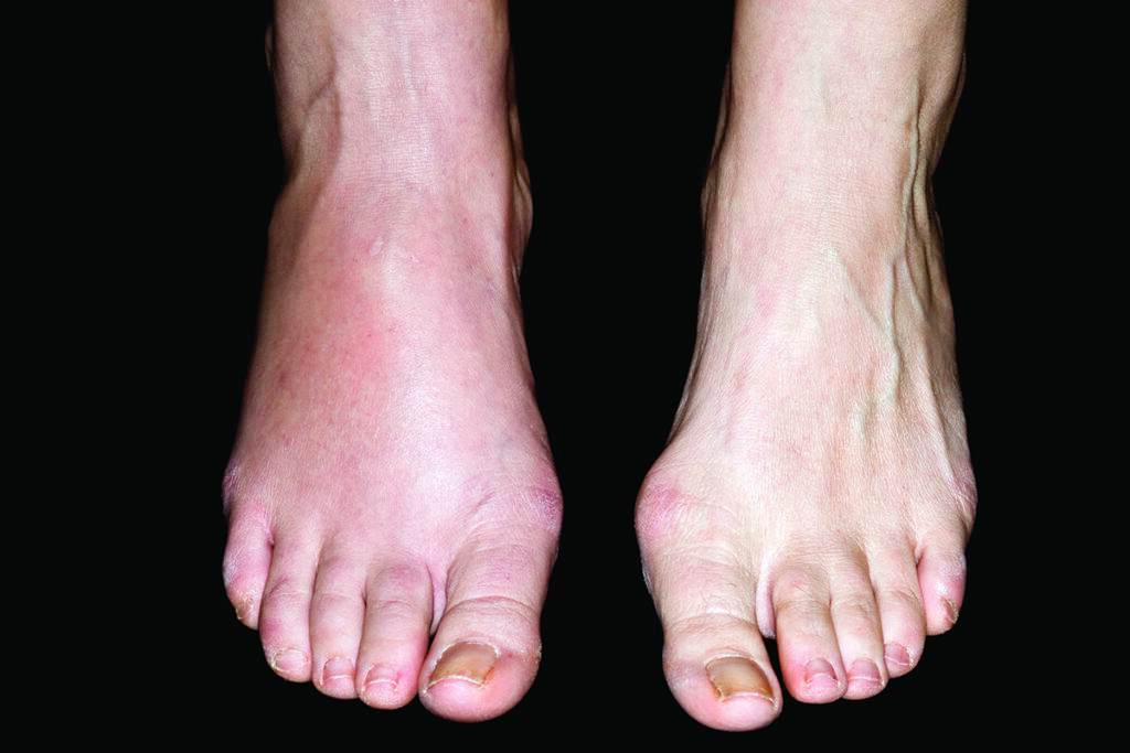
Case 1: Stress fracture in the foot
A 44-year-old recreational runner attends your clinic. ‘I’ve been training for the marathon, but the pain in my foot is becoming unbearable. It used to come on at the end of my run but now it starts after a mile. I can’t remember ever injuring it. What’s causing it?’
Stress fractures occur from repetitive submaximal loads being placed on bone, to the extent that bone resorption occurs at a faster rate than bone remodelling. This leads to a microscopic ‘crack’, which if not given time to heal will lead to macroscopic damage. Stress fractures account for 10-20% of all sports medicine consultations with elite track and field athletes.1 The large majority (95%) of these occur in the lower limb.2 Tibial shaft stress fractures account for half of lower limb stress fractures while metatarsal fractures account for 10-20%.3 Patients will be out of sport for up to 13 weeks after diagnosis so early management is crucial.4
Typical presentation
Stress fractures commonly occur in runners, athletes competing in pitch-based sports and military personnel.5 They can occur with increased load, for example an increase in running distance, or reduced recovery time between loading periods, for example through upping the number of running sessions per week.
Key history
It is vital to elicit the patient’s activity level and training factors, such as volume, intensity and activity surface. While stress factors are usually the result of excessive load in active patients, they can occur when normal loads are applied to weakened bones, for example in those with low bone density or osteoporosis. Other important risk factors include female sex, hormonal issues (indicated by amenorrhoea or dysmenorrhoea in women and erectile dysfunction and slow hair growth in men) and poor nutritional status (not taking in sufficient calories).6,7 These factors are part of the relative energy deficiency in sport (RED-S) condition.
Patients with a stress fracture will usually complain of pain that is insidious in onset and localised. It will be worse with activity and repeated loading and will subside with rest. Gradually, this pain will get worse and present earlier in a training session or start to be felt during daily activities. The patient will usually have no history of trauma. Any night-time pain is a red flag for an alternative diagnosis, and requires urgent investigation.
Examination
Palpate the affected areas for bony tenderness. Commonly stress fractures affect the tibia, navicular, fibula, metatarsals and calcaneus. There will be localised bony tenderness and possibly redness, swelling and bruising or warmth. Local tenderness is present in more than two-thirds of cases and oedema in around a fifth to two-fifths.8 It may also be helpful to ask the patient to perform graduated calf-raises or hopping to see if this reproduces the pain, which increases the likelihood of a stress fracture. If there is diagnostic uncertainty, you should also perform a full neurovascular examination to rule out alternative diagnoses.
Management
Radiological investigations are crucial if you suspect a stress fracture. Changes range from a mild periosteal reaction to a visible fracture line. However, X-ray changes occur late in the presentation and may not occur at all, so a normal X-ray does not exclude the diagnosis.9 If you have a high suspicion of a stress fracture and X-rays are normal, refer the patient for more sophisticated imaging, such as CT, MRI and bone scans and refer to orthopaedic surgery to confirm the diagnosis, start management and arrange follow-up.
Reassure patients that most stress fractures can be managed by activity modification. The key to successful treatment is a period of reduced loading. You may recommend non-weightbearing activities such as cycling and swimming to allow the patient to maintain aerobic fitness. Activity progression should be gradual, according to the patient’s level of pain. Any activity that leads to aggravation of symptoms should be avoided. With a gradual increase in intensity, the patient should be able to return to pre-injury activity levels. Stress fractures affecting the navicular and fifth metatarsal or that fail conventional treatment are at higher risk of non-union. These may require non-weightbearing cast immobilisation or surgery.
Consider risk factor modification, for example optimising bone health in those at risk of reduced bone mineral density, dietitian input for nutritional issues, podiatry input for biomechanical issues with running technique and referral to endocrinology for hormonal deficits and to investigate possible RED-S.
Differential diagnoses
It is important to rule out alternative diagnoses, such as medial tibial stress syndrome (‘shin splints’), peroneal nerve entrapment and chronic exertional compartment syndrome.
In the case of medial tibial stress syndrome, there is usually a dull, achy pattern of pain on the medial side of the tibia. There is a characteristic tenderness over the medial border of the tibia on examination, which is more diffuse than in a stress fracture. Also, unlike with a stress fracture, patients can usually hop repetitively on the affected leg without considerable pain. Peroneal nerve entrapment will cause paraesthesia over the antero-lateral calf and dorsum of the foot. The patient may complain of weakness or inco-ordination in the affected ankle or foot and on examination there will be weakness with dorsiflexion and eversion. Chronic exertional compartment syndrome is another differential diagnosis (see Case 2).
Case 2: Chronic exertional compartment syndrome
A 42-year-old competitive runner attends your practice. He tells you that since he’s had to start running on harder ground, he has had a burning pain at the bottom of both of his legs. ‘It’s so strange. I can only run for 15 minutes before the pain becomes too much. When I stop running, the pain goes away straight away. Do you think I have shin splints?’
Chronic exertional compartment syndrome (CECS) is an ischaemic condition that occurs when a fascial compartment cannot accommodate the increase in volume associated with muscle contraction and swelling, leading to an increased pressure. This is usually because of repetitive and strenuous exercise when increased blood flow and muscle volume cause small blood vessels to be compressed, resulting in reduced blood flow and ischaemia. It must be differentiated from limb-threatening acute compartment syndrome that complicates lower limb fractures, burns and drug overdoses.
The diagnosis of CECS is often delayed, partly because patients may be unable to explain the pain in specific terms and clinicians may not be aware of it. One study found CECS was the primary cause of chronic anterior leg pain in 14% of cases referred.10
The typical history
CECS manifests as aching, burning or cramping in the affected compartment and numbness, weakness and in severe cases, foot drop. There may be distal neurological symptoms of paraesthesia in the foot. The pain usually increases with continued exercise.
Classically, the pain is relieved when the activity is stopped and is bilateral 70-80% of the time.11
Differential diagnoses include medial tibial syndrome (shin splints), stress fractures and popliteal entrapment syndrome, which should be ruled out by other signs or investigations.
Examination
Absence of inner border tibial tenderness will rule out shin splints and should raise the suspicion of CECS. There is often little or nothing to see in CECS, however. There may be pain on palpation of the affected muscle compartment. Unlike popliteal entrapment syndrome, arterial pulses are usually intact as CECS primarily affects the venous system. If the examination is normal, ask the patient to exercise until they start to get symptoms and then re-examine them, looking for pain on palpation or with passive stretching, and a feeling of firmness of the muscles in the involved compartment.
Understanding the anatomy of the four lower leg compartments is key to diagnosing CECS.10
- Anterior – loss of sensation in first web space and weakness with ankle dorsiflexion.
- Lateral – weakness of eversion and numbness on the dorsum of the foot.
- Superficial posterior – numbness of the lateral foot and distal calf.
- Deep posterior – weakness of plantar flexion and numbness on the plantar aspect of the foot.
Management
If the history and symptoms are convincing, refer to orthopaedic surgery. Diagnosis requires intracompartmental pressure measurement, an invasive investigation that should only be performed if other diagnoses are excluded and the patient is considering surgical intervention.
You may consider starting conservative management straight away. Evidence shows early discontinuation or reduction of the eliciting activity, arch support through orthotics, and education on footwear and running surfaces are the most effective conservative options.12 Patients should be advised to change their running shoes every 500 miles and to avoid excessive running on hard surfaces.13
Case 3: Acute lateral ankle ligament sprain
A 23-year-old amateur football player sees you for an urgent same-day appointment. He explains his ankle ‘gave way’ during a match the night before. He tried to play on after it happened but is in a lot of pain today. He is keen to know when he will be able to play again, as he has an important cup match in two weeks.
Acute lateral ligament sprain is very common in sport and as a presentation to both GP practices and A&E. The typical mechanism of injury is an inversion force on a plantarflexed foot;14 the patient will often report that they ‘went over their ankle’ or ‘rolled their ankle’ when running or walking. The lateral ligaments of the foot include the anterior talofibular ligament (ATFL), the calcaneofibular ligament (CFL) and the posterior talofibular ligament (PTFL); the ATFL is the most commonly injured and the weakest of the three.14,15 The PTFL is the least commonly affected ligament, typically only ruptured in severe injuries such as ankle dislocation.
Examination
It is crucial to discern whether the patient has a lateral ligament injury or a more significant injury requiring immobilisation or referral to secondary care.
A functional assessment is a useful starting point. Simply asking the patient to stand on the inside and outside of their feet is an effective measure to gauge the subtalar range of motion and level of pain.
Palpate important landmarks such as the lateral malleolus, posterior aspect of distal fibula, ATFL, base of fifth metatarsal, talar dome, deltoid ligament, medial malleolus, navicular tuberosity and dorsum of navicular. Any tenderness should raise suspicion of injury in the respective structures. You should also palpate proximally up the syndesmosis and along the whole length of the tibia and fibula.
Then assess ankle movement and strength, including passive and resisted ankle dorsiflexion, plantarflexion, eversion and inversion. Pain with passive or resisted inversion would raise suspicion of a lateral ligament injury. Specialised tests include the anterior drawer test and inversion stress test.16
The Ottawa ankle and foot rules can help you to decide if referral for radiographic studies is needed: they can exclude the need for fractures of the ankle and mid-foot, demonstrating sensitivity of almost 100%.17 Refer for an ankle X-ray if there is any pain in the malleolar zone and:
- Bone tenderness along the distal 6cm of the posterior edge of the tibia or tip of the medial malleolus or
- Bone tenderness along the distal 6cm of the posterior edge of the fibula or tip of the lateral malleolus or
- An inability to bear weight.18
The Ottawa rules indicate a foot X-ray series if there is pain in the midfoot zone and:
- Bone tenderness at the base of the fifth metatarsal (for foot injuries) or
- Bone tenderness at the navicular bone (for foot injuries) or
- An inability to bear weight.18
- If the ankle has impaired proprioception, which may be the case with chronic ankle instability (CAI), they may find it difficult to stand on one leg whilst looking at the ceiling.
Management
Most acute lateral ankle sprains can be managed non-surgically. Initial immediate advice to patients should be to follow the RICE method – rest, ice, compression and elevation. Thereafter it should be tailored to the individual, considering the severity of the injury. Long-term immobilisation can be detrimental, but severe injuries benefit from short-term immobilisation.19 Patients should be advised about adequate analgesia and referred to physio for rehabilitation; in the meantime Alphabet exercises (drawing each letter of the alphabet with the ankle while eyes closed) will help get the ankle mobile within 24-48 hours of injury.
Time out of sport will vary and is difficult to predict. There is also a lack of formal advice on return to sport, but this should be based on testing functional performance as part of a rehabilitation program, including comparison with the contralateral side and testing agility.20
How to approach chronic ankle instability
Around 10-30% of patients with acute lateral ligament sprains go on to develop CAI, which occurs with repeated or inadequately managed ankle sprains.21 Patients typically describe the ankle ‘giving way’, particularly on uneven surfaces.
CAI includes both mechanical and functional instability related to recurrent ligamentous injury. Mechanical instability involves anatomical changes including increased ligament laxity, whereas functional instability involves persistent symptoms despite a lack of objective evidence on examination of ligament laxity. This type of instability is typically due to proprioceptive impairment and reduced neuromuscular control. Risk factors include an underlying foot deformity, postural imbalance, raised BMI and a history of previous significant ankle injury.
If the ankle has impaired proprioception, the patient may find it difficult to stand on one leg whilst looking at the ceiling.
Players with CAI should complete a 10-week physiotherapy-led proprioceptive rehabilitation programme. If the patient aims to continue competing in sport, they should see a physiotherapist to ensure the ankle is adequately taped to prevent re-injury. If instability remains after an intensive 10-week proprioceptive programme, refer to orthopaedic surgery for consideration of surgical stabilisation of the lateral ligaments.
Dr Neil Heron is a GP and consultant in sports medicine at Queen’s University, Belfast, and Dr Dáire Rooney and Dr Emma Gilmartin are FY2 doctors with an interest in sports medicine at the Royal Group of Hospitals, Belfast
References
- Bennell K, Malcolm S, Thomas S et al. The incidence and distribution of stress fractures in competitive track and field athletes. A twelve-month prospective study. Am J Sports Med 1996;24(2):211-217
- Liong S, Whitehouse R. Lower extremity and pelvic stress fractures in athletes. Br J Radiol 2012;85(1016):1148-1156
- Hulkko A, Orava S. Stress fractures in athletes. Int J Sports Med 1987;8(3):221-226
- Miller T, Jamieson M, Everson S et al. Expected time to return to athletic participation after stress fracture in division I collegiate athletes. Sports Health 2018;10(4):340-4
- Tuan K, Wu S, Sennett B. Stress fractures in athletes: risk factors, diagnosis, and management. Orthopedics 2004;27(6):583-593
- Wright A, Taylor J, Ford K et al. Risk factors associated with lower extremity stress fractures in runners: a systematic review with meta-analysis. Br J Sports Med 2015;49(23):1517-1523
- Bennell K, Matheson G, Meeuwisse W et al. Risk factors for stress fractures. Sports medicine 1999;28(2):91-122
- Patel D, Roth M, Kapil N. Stress fractures: diagnosis, treatment, and prevention. Am Fam Physician 2011;83(1):39-46
- Wright A, Hegedus E, Lenchik L et al. Diagnostic accuracy of various Imaging Modalities for Suspected Lower Extremity Stress Fractures: A systematic review with evidence-based recommendations for clinical practice. Am J Sports Med 2016;44(1):255-263
- Blackman P. A review of chronic exertional compartment syndrome in the lower leg. Med Sci Sports Exerc 2000;32(3 Suppl):S4-10
- Raikin S, Rapuri V, Vitanzo P. Bilateral simultaneous fasciotomy for chronic exertional compartment syndrome. Foot Ankle Int2005;26(12):1007-1111
- Cook S, Bruce G. Fasciotomy for chronic compartment syndrome in the lower limb. ANZ J Surg 2002;72(10):720-723
- Rethnam U, Makwana N. Are old running shoes detrimental to your feet? A pedobarographic study. BMC Res Notes 2011;4(1):1-5
- Al-Mohrej O, Al-Kenani N. Acute ankle sprain: conservative or surgical approach? EFORT Open Rev 2016;1(2):34-44
- Ferran N, Maffulli N. Epidemiology of sprains of the lateral ankle ligament complex. Foot Ankle Clin 2006;11(3):659-962
- Wolfe M, Uhl T, Mattacola C et al. Management of ankle sprains. Am Fam Physician 2001;63(1):93-104
- Bachmann L, Kolb E, Koller M et al. Accuracy of Ottawa ankle rules to exclude fractures of the ankle and mid-foot: systematic review. BMJ 2003;326(7386):417
- Stiell I, McKnight R, Greenberg G et al. Implementation of the Ottawa Ankle Rules. JAMA 1994;271(11):827-832
- McGovern R, Martin R. Managing ankle ligament sprains and tears: current opinion. Open Access J Sports Med 2016;7:33-42
- Plisky P, Rauh M, Kaminski T et al. Star excursion balance test as a predictor of lower extremity injury in high school basketball players. J Orthop Sports Phys Ther 2006;36(12):911-919
- Vuurberg G, Hoorntje A, Wink LM et al. Diagnosis, treatment and prevention of ankle sprains: update of an evidence-based clinical guideline. Br J Sports Med 2018;52(15):956
Visit Pulse Reference for details on 140 symptoms, including easily searchable symptoms and categories, offering you a free platform to check symptoms and receive potential diagnoses during consultations.