The non-fungal annular rash – how to identify granuloma annulare

Under the radar: GP and dermatology specialist Dr Anjali Pathak describes how a case of granuloma annulare was initially treated as a fungal infection, and explains how it can be identified and managed promptly
Note this case is hypothetical and developed for educational purposes
Clinical history
A 34-year-old woman presents with a slowly enlarging ring-shaped lesion on the dorsum of her right hand. It has been present for around six months and has gradually become more noticeable. It is asymptomatic, with no itch, pain or tenderness. She is otherwise well, with no systemic symptoms.
She initially assumed it was ringworm and tried over-the-counter topical clotrimazole and terbinafine for several weeks, with no improvement. She then saw her GP and was prescribed a combination preparation containing miconazole and hydrocortisone. Despite this, the lesion continued to enlarge slowly. She is worried that she has a persistent fungal infection and asks whether she needs a stronger antifungal.
On examination, there is a smooth annular plaque on the dorsum of her right hand, made up of small, firm, skin-coloured to pink-brown papules. There is no surface scale, vesiculation, crusting, erosion or scaly advancing edge. When the border is palpated, it feels raised and papular.
The clinical diagnosis is localised granuloma annulare.
Making the diagnosis
The key clue is the lack of scale (and, in this case, lack of response to anti-fungals). Granuloma annulare is usually smooth, with a papular edge. Tinea corporis tends to have scale at the active edge and is often itchy. Palpation can help, as granuloma annulare may feel like a ring of small dermal papules rather than a flat, scaly plaque.
Granuloma annulare is a benign inflammatory skin condition. It affects all ages, but localised disease is most often seen in children and young adults, with a female predominance. It commonly affects the backs of the hands and feet, fingers, ankles, elbows and other extensor sites (see image).
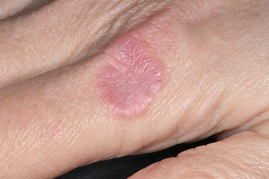
Image: Granuloma annulare on the hand of a 52 year old female patient
Lesions often start as small, firm papules that gradually spread outwards to form annular or arcuate plaques. They may be skin-coloured, pink, violaceous or brown, depending on skin tone and stage. In skin of colour, erythema may be subtle, so lesions may look more brown or violaceous than red.
The cause is unclear. Granuloma annulare appears to represent a delayed inflammatory or hypersensitivity-type reaction in the dermis. Minor trauma, infections and insect bites have been described as possible triggers, although many patients have no obvious precipitant.
Most cases seen in general practice are localised. Less common forms include generalised granuloma annulare, which can be more persistent and widespread; subcutaneous granuloma annulare, usually seen in children as deeper rubbery nodules; and perforating granuloma annulare, which may show crusting or umbilicated papules.
In a typical localised case, the diagnosis is clinical. If fungal infection remains a realistic possibility, take skin scrapings before prescribing another antifungal. Other differentials depend on site and context and may include discoid eczema, psoriasis, lichen planus, erythema migrans, sarcoidosis and necrobiosis lipoidica. Patients can be referred to secondary care where there is uncertainty regarding diagnosis.
Granuloma annulare has reported associations with diabetes, dyslipidaemia and thyroid disease. Disseminated granuloma annulare has also been linked with HIV. Further investigations should be considered where disease is generalised, recurrent or unusually persistent, or where there are relevant symptoms or risk factors.
How should it be managed?
Reassurance is usually the most important part of management. Patients are often relieved to hear that this is not ringworm, not contagious and not dangerous. Most localised cases do not need active treatment. Around half resolve within two years, although lesions can last longer and recurrence can occur.
For persistent or cosmetically troublesome lesions, treatment options include a potent topical corticosteroid, intralesional corticosteroid for a small persistent lesion, or a topical calcineurin inhibitor such as tacrolimus or pimecrolimus off-license. Cryotherapy is an option, but should be used cautiously because of the risk of scarring or pigmentary change, particularly in skin of colour.
Widespread or refractory disease can be referred to dermatology. Phototherapy or systemic treatments such as hydroxychloroquine, methotrexate or short courses of systemic corticosteroids may be considered in secondary care, but responses are variable.
In this case, the patient chose observation after reassurance. She was advised to return if the rash became scaly, painful, ulcerated, rapidly progressive or widespread.
Learning points for GPs
- Consider granuloma annulare when an annular lesion is smooth rather than scaly. Palpation can help, as the edge may feel raised and papular.
- If fungal infection remains possible, take skin scrapings before prescribing repeated empirical antifungal treatment.
- Most localised cases can be managed with reassurance. Explain that granuloma annulare is benign, inflammatory and not contagious, but may take months to years to resolve.
- Consider further investigations if disease is generalised, recurrent or unusually persistent, or if there are relevant symptoms or risk factors. Seek dermatology advice if lesions are atypical, widespread, ulcerated, rapidly progressive or diagnostically uncertain.
Dr Anjali Pathak is a specialty dermatology physician and GPwER in dermatology based at West Hertfordshire Teaching Hospitals NHS Trust
Sources and further reading
- DermNet: Granuloma annulare. Last reviewed February 2025
- Primary Care Dermatology Society (PCDS). Clinical guidance: Granuloma annulare. Last updated February 2024
- Joshi T, Duvic M. Granuloma annulare: An updated review of epidemiology, pathogenesis, and treatment options. Am J Clin Dermatol 2022 Jan;23(1):37-50
Visit Pulse Reference for details on 140 symptoms, including easily searchable symptoms and categories, offering you a free platform to check symptoms and receive potential diagnoses during consultations.
Related Articles
READERS' COMMENTS [1]
Please note, only GPs are permitted to add comments to articles


Useful and concise article, thank you.